Acetabular Fractures San Francisco

The normal hip is a “ball and socket” type joint. The ball is called the femoral head, the socket is called the acetabulum. The normal movement of the hip depends upon the two surfaces fitting together precisely. The two surfaces are coated with hyaline cartilage which is a very low friction surface, smoother than even the best man made surface.

AP radiograph 2 years after open reduction internal fixation of anterior column plus posterior hemitransverse acetabular fracture in a 65 year old weekend warrior. This patient has returned to cycling and sporting activity.

When a fracture of the hip joint occurs, commonly it involves the femur or ball side of the joint. Rarely if forces are severe enough, the fracture injures the socket. This may occur as the femoral head is forcefully driven into the acetabulum. Sometimes the hip dislocates as the acetabulum fractures.

These injuries generally occur as the result of high energy injuries, such as a fall from heights, automobile or motorcycle accidents, bicycle crashes, and other forceful injuries of the hip joint.

High energy injuries of the hip are commonly associated with other injures and problems. The fractured bony surfaces and surrounding injured soft tissues lead to internal bleeding. Major blood vessels may be lacerated or torn. The sharp fractured surfaces can damage the bowel or bladder. The nerves that provide sensation and motor function to the leg and foot can be injured. In some instances the nerves that control bowel and bladder function or sexual function can be injured by the trauma as well.

The fractures generally fall into categories of patterns which were put into a classification system which generally dictates the approach to surgery. This classification is called the Letournel Classification and is presented below. (Figure 1)

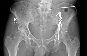
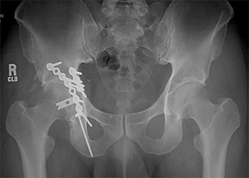
The result of these fractures is disruption of the smooth, congruent surface of the acetabular bone. Stepoffs or gaps in the position of the bones prevent the smooth articulation of the hip joint. If the bone heals with these irregularities, damage of the cartilage and painful arthritis of the hip is a predictable result. This usually results in severe functional limitations, and significant pain.

How does Acetabular Fracture Surgery work?

For the majority of patients, surgery is required to restore function to the hip. The goal of surgery is accurate reconstruction of the fractured bone. The pieces of the acetabulum are precisely placed back into position and secured with screws and/or plates. This reconstruction restores the smooth surface of the acetabulum and its accurate fit to the femoral head. This procedure is called “open reduction and internal fixation” or “ORIF” for short.

In the majority of cases, approximately 80%, arthritis can be prevented and there is normal or near normal hip function after the surgery.

AP radiograph 2 years after open reduction internal fixation of posterior column plus posterior wall acetabular fracture in a 39 year old competitive athlete. This patient has returned to playing American football.

Prior to surgery, patients undergo a comprehensive evaluation of their medical stability. This data is essential to the surgeon, medical specialists, anesthesiologist and other members of the health care team. X-rays are also obtained, including 5 standard pelvis views and CT (computed tomography) scans. Sophisticated computer software allows the technologist to make cross-sectional CT scans into three-dimensional images. These x-rays enable the surgeon to determine the fracture pattern and precise degree of displacement and to plan the surgical approach.

For best results, surgery should take place between two and seven days of injury. The potential for difficulties and complications increases after three weeks.

Depending on the fracture pattern, usually one of the following three surgical approaches will be chosen which will give the best access for reconstruction of the acetabulum:

  • Kocher-Langenbeck approach (posterior approach)
  • Ilioinguinal approach (simultaneous access to both anterior & posterior portions of the pelvic ring)
  • Extended iliofemoral approach (lateral approach)

The patient is placed on a special operating table that applies traction to the leg during surgery and assists in reducing the fracture.

The operation itself takes between two and five hours, with a blood loss ranging from 250 to 2,000 cc’s. A blood transfusion may be necessary during or after surgery. A device will be utilized during surgery to collect blood lost by the patient and to return it to their circulation. During surgery, drains will be placed in the surgical site, which will be removed two to three days post-operatively. The dressing covering the incision will be checked on a regular basis and changed or removed two to three days after surgery. Patients wear compression boots and anti-embolus stockings to prevent blood clots from forming in the large veins or veins traveling to the lungs.

What are possible complications?

Surgical wound infection and injury to nerves are the most common significant complications. The chance of either one is about three percent. Another rare complication is injury to a major blood vessel or other internal organ.

If an infection develops, the surgical wound is cleaned in the operating room and an antibiotic is given intravenously. In most cases, infections have little effect on function other than prolonging hospitalization and recovery. Some infections can impair function by damaging the joint cartilage.

Pressure on nerves during surgery can cause impaired nerve function. Gaining access to the bone during surgery requires the surgeon to pull nerves gently aside. A foot drop is the most common problem though usually resolves within months after the surgery.

How long is the recovery for acetubular fracture surgery?

Patients spend the two hours following surgery in the recovery room where nurses closely monitor them. The first night after surgery is typically spent in the intensive care unit to facilitate close monitoring and after that, the patient is returned to the normal hospital ward.

The same team of medical specialists cares for each patient pre- and post-operatively. Included among the team’s post-surgical priorities are pain management, preventing infection, and the prevention of deep vein thrombosis (blood clots in large veins), and pulmonary embolus (blood clots traveling through veins to the lungs).

Patients begin physical therapy as soon as possible to improve hip motion and muscle function and to learn to use appropriate assistive devices such as crutches or a walker. During the first eight weeks following surgery, the injured hip should bear no more than a limited weight of 30 pounds. Placing full weight on the injured side prior to bone healing can cause the screws or plates which hold the broken bone together to bend or break. Too vigorous exercise such as resistive exercise against weights can also cause failure. If failure occurs, re-operation may be necessary and the chance of developing arthritis is greatly increased.

Discharge from the hospital usually occurs within seven to ten days of surgery, depending on surgical wound healing, progress in physical therapy and complications. At discharge, medication is prescribed for pain. Patients may also be given an antibiotic to prevent or treat infection, an anticoagulant to prevent blood clots, and medication to prevent ectopic, or abnormal bone formation.

After Discharge

Discharge from the hospital usually occurs within seven to ten days of surgery, depending on surgical wound healing, progress in physical therapy and complications. At discharge, medication is prescribed for pain. Patients may also be given an antibiotic to prevent or treat infection, an anticoagulant to prevent blood clots, and medication to prevent ectopic, or abnormal bone formation.

Pain and discomfort after discharge is natural considering the physical trauma patients have endured. The pain will increase and decrease at different times and affect various areas of the body. Dull pain is common during the first couple of weeks, and patients might also hear an occasional click or “pop” in the joint. New-numbness and a tingling sensation are common around the incision area. However, patients experiencing severe and consistent pain and/or discoloration of any kind should consult with the doctor.

In most cases patients should continue regular therapy with a physical therapist to regain motion and muscle strength. With compliance, a patient could show as much as 80% recovery at 6 months post surgery and a full recovery at one year. Follow-up outpatient visits are also necessary to allow progress to be monitored by x-ray and physical examination. Patients will be advised during these visits about future care and what physical activities are recommended.

Potential complications associated with acetabulum fractures include post-traumatic arthritis, ectopic bone formation, and avascular necrosis of the femoral head (lack of blood supply that will cause the bone to die).

Total Hip Replacement, an operation in which the hip joint is replaced with an artificial joint, is often used to repair damage caused by arthritis. This is often a good solution for arthritis, but in the context of an acetabular fracture, commonly the fracture needs reduction and fixation prior to being able to support an implant.